As we covered the vertebrae in the previous post in the skeletal series, we shall move on to the last elements in the axial skeleton (bar the clavicle and scapula in next post). The elements of the appendicular skeleton will follow shortly.
Excavation
As before, great care must be taken when excavating the ribs. I have carried out micro-excavation of juvenile remains, (when I volunteered at Humber Field Archaeology) that consisted of the vertebrae, half of the ribcage and parts of the pelvis in-situ, and it took a while. During excavation of human remains on site, it is very unlikely that the rib cage will be in its natural anatomical position. Due to the soil and weight of the earth above the body, and the movement in the intervening period between burial and excavation, the ribs are likely to be broken and misplaced. As always keep a look out for finer skeletal finds.
The Rib Cage
The focus of this post will be the sternum, which is made up of the Manubrium, Corpus Sterni (or Gladiolus) and the Xiphoid Process alongside a look at the ribs, all of which help to form the rib cage. It is necessary to note here that variation in the number of ribs, as of the vertebrae discussed previously, can differ in people and in archaeological populations (Mays 1999: 15). The function of the rib cage, as the main upper part of the torso in the human body, is to protect the vital organs that lie within the protective enclave of the ribs. They include the majority of the torso organs such as the heart, liver, lungs, kidneys and partially the intestines. The rib cage also helps breathing by the function of the intercostal muscles lifting and lowering the rib cage, aiding inhalation and exhalation. The ribs are attached dorsally to the vertebrae, with articulating facets for the tubercle end of the ribs for the thoracic vertebrae (White & Folkens 2005).
Rib Cage Anatomy, Terminology and Elements
The number of ribs present in the typical human skeleton is of 12 paired rib elements (a total of 24 altogether). Ribs project from proximal articulating facets with thoracic vertebrae, slant forward, and depending on the rib pair under consideration, articulate at the distal end with either the sternum, hard cartilage or ‘float’ freely (Jurmain et al 2011). Ribs usually increase from size from rib 1 to rib 7, and decrease in size from rib 7 to rib 12 (White & Folkens 2005: 185).
The Upper 7 ribs on each side of the cage connect distally directly to the sternum via cartilage, whilst the 8th, 9th and 10th ribs connect indirectly to the sternum. The last 2 ribs are often called ‘floating’ ribs because they have ‘short cartilaginous ends that lie free in sides of the body wall’ (White & Folkens 2005: 181).
The basic landmark anatomy of a rib includes the head, neck, tubercle which articulates with the thoracic vertebrae & the long shaft of the rib. In the picture below the head of the ribs are medial whilst the sternal ends are lateral. The side that is superior is called the cranial edge, whilst the inferior is called the caudal edge. It can be fairly easy to side a loose or partial rib as the Cranial edge is fairly thicker and blunter when compared to the grooved and sharp caudal edge (White & Folkens 2005: 192).

The rib cage laid out from 1st to 12th ribs, note the size and shape morphology. Occasionally an individual will have only 11 paired ribs or may have an extra pair, this is natural variation. (Image credit: Shutterstock).
Distinctive Rib Cage Elements
- The 1st rib is most unusual and can usually be identified the easiest. It is particularly blunt, broad and thick, as well as this it has no caudal groove (Roberts & Manchester 2010).
- The 2nd rib serves as an intermediary to the 1st and 3-9th more regular ribs. It has a large tuberosity for the serrator anterior muscle half way along its length (White & Folkens 2005: 187).
- The 11th rib lacks a tubercle and the sternal end is often pointed.
- The 12th rib is shorter then the 11th rib and may be even shorter then the 1st rib. It lacks the angle and costal groove, and is often easy identifiable (May 1999).
Sternum
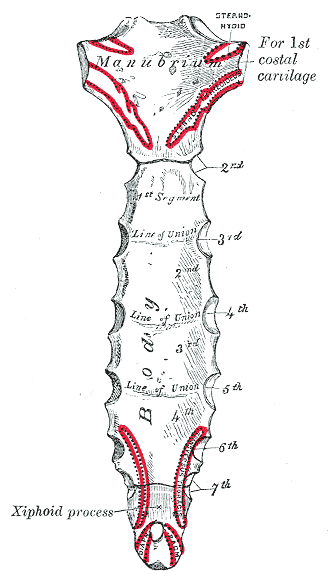
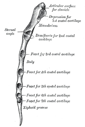
As stated the sternum is made of 3 individual bones, those of the manubrium, corpus sterni and the Xiphiod Process. As made clear by the diagrams below, there are 7 facets located laterally for the anterior of the ‘true’ ribs alongside the corpus sterni and manubrium. The sternum is composed of these three elements in adulthood but develops from 6 segments (White & Folkens 2005: 181).
The manubrium is the thickest and squarest part of the sternum bones and should be easily identifiable as such. At the superior corners there are clavicular notches located, which articulates with the right and left clavicles. The clavicles and scapula help to form the shoulder girdle and will be discussed in the next post.

The three individual elements of the sternum, with the manubrium (proximal), corpus sterni (centre) and xihoid process (distal) highlighted. (Image credit: Wikipedia 2011).

Lateral view of sternum elements with the individual rib facets highlighted. (Image credit: Wikipedia 2011).
The corpus sterni is rather thin in comparison to the manubrium, and it is often said to be ‘bladelike’. Again, costal notches are present as seen in the above picture. They cater for ribs 2-7 (Mays 1999).
The xiphoid process can be found inferior to the corpus sterni but, depending on the age of the person involved, may not be found in archaeological samples. The process shares the seventh costal notch. This bone is often highly variable in shape, and is late to ossify (White & Folkens 2005: 184).
A Pre-contact Peruvian Case Study
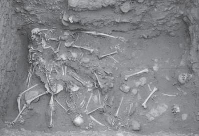
The site of Pacatnamu, in the Jequetepeque River valley on the northern Peru coast, provides a site where mutilated human remains and contextual information has been unearthed (Larsen 1997: 137). At this Moche site (100-800 AD), evidence has been found of executed captives who were thrown into a trench at the bottom of an entrance to a ceremonial precinct (Verno 2008: 1050). The skeletal group found & studied was composed of 14 adolescent and young adult males, who were recovered from 3 superimposed layers at the site. The superimposed layers were indicative of 3 distinct burial episodes (Larsen 1997: 137). From the evidence on the skeletal elements, it seems that weathering took place after death but before burial. The evidence is backed up by the palaeoenvironmental remains of the presence of the pupal cases of muscoid flies (Verano 1986).
It seems that the display of the decomposing bodies, together with a lack of a proper burial, was clearly intentional. In the topmost layer of the burial episodes, multiple stab wounds were found on both the vertebrae and rib elements. This pattern is broken by the bottom and middle layer where the pattern is more towards decapitation or throat slashing as evidence by cutmarks on the cervical vertebrae (Larsen 1997: 137). Of particular interest is the evidence of five individuals from the middle and lower deposits that have bisected manubriums with evidence of fractured ribs, which is suggestive of the chest cavity being opened forcibly (Verano 1986).
Larsen (1997: 137) remarks that on the ‘basis of the age distribution, sex, and evidence of healed and unhealed injuries (rib fractures, depressed cranial fractures), Verano (1986) speculates that they were war prisoners’. The conclusion is well supported from the cultural representations in the art and architecture of the Moche culture. Many cultures throughout the world made, and continue to make, sacrifices of various kinds; in particular it is thought that for pre-contact South American cultures human sacrifice represented ‘the most precious form of sacrifices, and seems to have been reserved for particularly important rituals and events’ (Verano 2008: 1056).
It has also been noted that the capture and killing of enemies was a common practise in communities in pre-contact South America. Such killings can also incur within ritual presentations and displays. There are many such examples throughout South American, and indeed throughout the Americas (Teotihuacan, Punta Lobos, Sipan) (Verano 1986). A careful consideration through the integrated studies of the archaeological sites, bioarchaeological study of the human material, together with careful ethnographic comparisons, can help to understand the processes and results of human sacrifice in South American cultures.
Further Information
- This website here gives a quick and easy run down of the features of the rib cage.
- For a detailed look at how ribs and vertebrae articulate visit this site or this site.
Bibliography:
Jurmain, R. Kilgore, L. & Trevathan, W. 2011. Essentials of Physical Anthropology International Edition. London: Wadworth.
Larsen, C. 1997. Bioarchaeology: Interpreting Behaviour From The Human Skeleton. Cambridge: Cambridge University Press.
Mays, S. 1999. The Archaeology of Human Bones. Glasgow: Bell & Bain Ltd.
Roberts, C. & Manchester, K. 2010. The Archaeology of Disease Third Edition. Stroud: The History Press.
Verano, J. W. 1986. ‘A Mass Burial of Mutilated Individuals at Pacatnamu‘. In C. B. Donnan & G. A. Cock. (eds.) The Pacatnamu Papers. 1. pp.117-138. Los Angeles: Museum of Cultural History, University of California.
Verano, J. W. 2008. ‘Trophy Head-Taking and Human Sacrifice in Andean South America‘. In H. Silvermann & W. Isbell.(ed.) Handbook of South American Archaeology. pp.1045-1058. Los Angeles: Springer.
White, T. & Folkens, P. 2005. The Human Bone Manual. London: Elsevier Academic Press.


Hi I’m a 52 year old man use to be a short 5 ft 6 inches tall but always looked a lot shorter and lousy body proportion genetics. Small boned with tiny torso abnormal low shoulder slanted dowown about 11 inches from shoulder to elbow Same.small forarms wrist and nands not broad at all with a 19 -nch leg. Skinney head now and aging rapidly 27 – 28 inseam. 4 ft 4 ft tall at 13. Abnormal. 9 inch shoe. Confusing should have been on hgh as a kid. My daughter at 5 ft inces tall has great high boned broad long torso looks 5 ft 6 longer arms then me my reach 64 -65 inch reach. Every one I see even kid at 6 – 8 yrs old look like great proportion genetics like adults just sorter I inherheted lousy genetics I always looked deformed face getting thinner and sickley with abnormal freaky hair loss all over scalp Andd spread out all loss of pigment propbably due to medication drugs. I’ve never seen hair like thiner then sowing thread it won’t even grow long in back all falling out everywhre. Its sickley not mail pattern baldness won’t even connect to eares 1 50th in diameter then Human hair. I. Have. Osteopenia 3 years ago maybe osteoporosis now ,ow energy. Always weird body little proportions. I can see my rib cage and only see 4 ribs under chest not 10 or 12 like you show , unless they go higher then chest how could I only have 4 ribs And you show 10 or 12 ? Is that abnormal ? Tnanks. Mitch.
Hi Mitch, thanks for visiting the blog. I’m sad to hear you have osteoporosis, I hope you remain in good health and take the necessary precautions!
Any individual human can be born with more or less then 12 ribs. 12 ribs is the standard number people are born with but there can be deviations. I think it is highly unlikely that you only have 4 however, as ribs are necessary in helping you to breath in and out, alongside helping toprotect the interal organs in the torso. If you take your right or left hand and feel down the side of your chest you can feel the larger ribs. it is very unlikely youll be able to feel the first 2 or 3, and the last 2 or 3 however as they are small and well covered by muscles. If you contunie to be worried I would consult your local doctor to get checked out! I hope this helps…
Hi. My 17 year old son said he just woke up one day and noticed his ribs appear deformed. He says the bottom one on the left is like dented in a bit. He says he is not in pain and can still do everything normally. Just says he looks deformed. I noticed he had a bad posture (lying on the side while on the computer) and wonder if he has squished his ribs from constantly being in this position. Do you have any ideas what this could be?
Hello Judy,
I would go see his Doctor and have a talk with him, just in case. Remember that at 17 years old the human skeleton, especially the male skeleton, is still growing. This could be a temporary cause of that. I would think it would be unlikely due to lying on the side whilst he is on the computer; that might be a short term effect in the body ‘shifting’ but after he gets up and moves around I’d imagine it’ll go back to normal. It takes a long concentrated effort for skeletal shape to due change due to forced pressures.
So please, if it still seems out of shape after a few hours, visit a doctor. Hope that helps, cheers for reading!
Very good and helping
Hi, I found a rib bone in my back yard. Is there an easy way to distinguish whether it is human or animal? ie sheep or dog? It is approx 30cm and not flat. Thanks
Hi Nicky,
It is probably best if you take a picture and email it to me so I can help! does the rib have either end piece? 30cm sounds quite large!
indeed clear and visible image illustration.And also well detailed and documented.it really helped and meet my research need. Thanks.
Thank you for reading!
I have a genetical extra rib on my left lower side of my rib cage. It is genetical as my great grandfather had it and all my 4 sons have it to. I have left my body to medical science at manchester university in event of death hopefully doctors and students may learn from this.
17/06/2016.
Hi Bryan,
That is wonderful and such a generous donation to make to the University of Manchester. I really appreciate your help and selfishness!
Yours,
David
Also, your email address is on the site so I removed it in case you receive a load of spam mail. I’ve got it still from the comment box you filled in. Thanks once again, and thank you for your interest in the site!
What is the reason that why last two ribs do not extend anteriorly ?? Although these are floating ribs !!
Hi Fareeha, I’m not sure! It just seems to be the shape and size of the floating ribs – perhaps they are an evolutionary trade for the flexibility of the torso/spine?